재생 버튼을 누르시면 해당 내용을 음성으로 들으실 수 있습니다.
습성 연령관련 황반변성 진단을 받은 후, 안과를 방문하면 광간섭단층영상(OCT) 검사를 진행합니다1. 광간섭단층영상(OCT) 검사는 눈 뒷부분을 촬영하는 비침습적인 진단 방법으로 삼출물의 유무 등 질환 상태를 확인할 수 있습니다1.
해당 검사를 통해 확인하는 두 가지 주요 삼출물은 망막내액(IRF)과 망막하액(SRF)입니다3. 눈에 이런 삼출물이 축적되면 시야가 흐릿해 보이거나 물결쳐 보이는 등 중심시력이 왜곡될 수 있습니다2, 3.
습성 연령관련 황반변성 환자의 광간섭단층영상(OCT) 검사 예시:
진료 때마다 정기적으로 광간섭단층영상(OCT) 검사를 진행해, 삼출물을 확인합니다1.
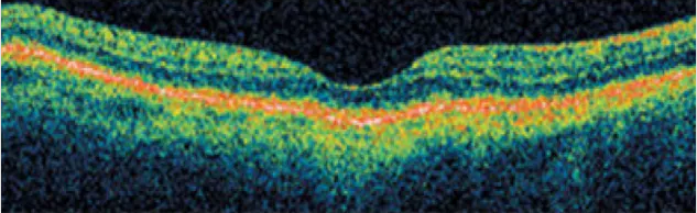
효과적인 치료가 이루어진 경우에는 눈 뒷부분에 삼출물이 거의 또는 전혀 없는 상태를 보입니다4.
아래 광간섭단층영상(OCT)에서는 눈 뒷부분에서 삼출물이 발견되지 않았습니다:
습성 연령관련 황반변성의 진행 상황을 확인하는 방법에 대해 더 알아보려면 여기를 클릭하세요.
References
1Kang SW, et al. The correlation between fluorescein angiographic and optical coherence tomographic features in clinically significant diabetic macular edema. Am J Ophthalmol 2004;137(2):313-322.
2. National Eye Institute. Facts About Age-Related Macular Degeneration. Available at https://nei.nih.gov/eyedata/amd. Accessed November 2019.
3Arnold J et al. The role of sub-retinal fluid in determining treatment outcomes in patients with neovascular age-related macular degeneration--a phase IV randomized clinical trial with ranibizumab: the FLUID study. BMC Ophthalmol. 2016;143(4):679-680.
4García-Layana A, et al. Early and intermediate age-related macular degeneration: update and clinical review. Clin Interv Aging. 2017;12:1579–1587.